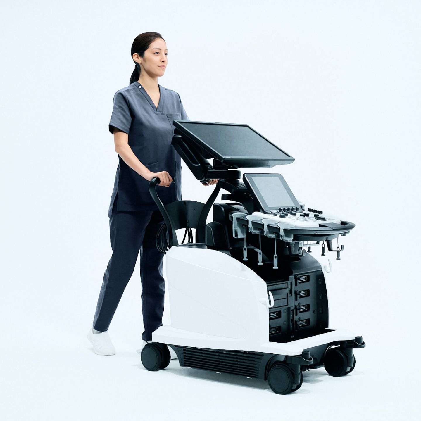
Dive deeper. See more. Decide with confidence.
DeepInsight x
 The new milestone of diagnostic ultrasound designed to deliver clarity, consistency, and confidence in every examination.
When clarity improves, confidence follows
DIx Solutions That Work as One
Powered by DeepInsight Fujifilm AI technology, The DeepInsight x elevates diagnostic ultrasound by combining advanced image intelligence, optimized system performance, and workflow automation so clinicians can focus on interpretation, not adjustment.

Deeper Image Intelligence

Uniform Clarity at Every Depth

Workflow That Adapts in Real Time
What sets DIx apart ?
A system designed around diagnostic confidence
Clearer images: Across the entire field. DIx delivers consistently high visibility from near field to far field, reducing blind spots and supporting wide-area observation without compromise.
Less manual adjustment, more consistency: Real-time optimization technologies adapt as the scanning plane changes, helping maintain image uniformity and reducing operator workload.
Advanced intelligence, applied practically: AI-based technologies are used where they matter most: improving image clarity, streamlining workflow, and supporting reproducible examinations always under clinician control.
See subtle structures. Preserve natural tissue expression.
Image quality that works with you
DIx is engineered to extract more meaningful information from every signal—without sacrificing natural image appearance.
Richer gradation and contrast help reveal subtle differences in tissue. Uniform image extraction supports confident assessment across the entire screen.Structure-enhancing processing highlights boundaries while preserving texture.
Detect what was previously difficult to see.
Flow visualization beyond conventional limits.
DIx introduces enhanced microvascular flow visualization that balances sensitivity and noise suppression. Low-velocity blood flow can be displayed with higher visibility, even in challenging conditions, helping support:
Colour Flow
eFlow
Detective Flow Imaging
All while minimizing motion-related artifacts that can obscure interpretation.
Experience streamlined workflow efficiency that frees up your time, letting you focus more on what matters most, delivering exceptional patient care
Automatic view recognition
Relevant body and probe markers are set automatically after image freeze, reducing repetitive manual input.
Real-time image optimization
Gain balance adapts dynamically as anatomy or scanning planes change—helping maintain consistency without constant adjustment.
Protocol-driven support
Pre-configured workflows and reference views help standardize examinations, support training, and reduce inter-operator variability.
shorter exams, smoother operation, and more predictable outcomes.
FUJIFILM's DIx Portfolio
Workflow that adapts to the examination
DIx is designed to reduce unnecessary interaction—so examinations flow naturally.


Move beyond visual estimation
Quantitative insight for liver assessment
DIx supports advanced, quantitative tools for liver disease assessment—helping clinicians evaluate steatosis and stiffness with greater objectivity.
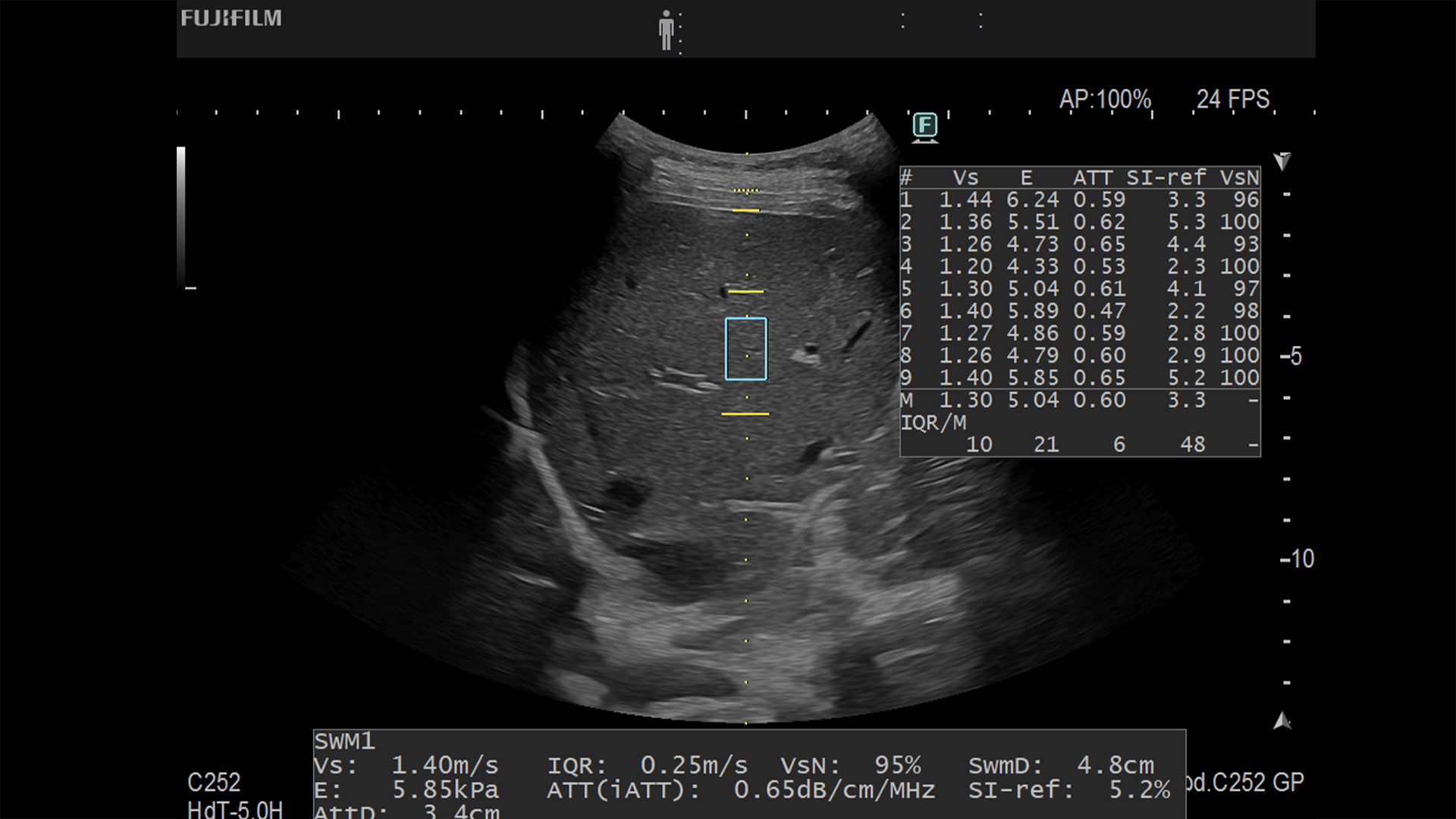
Shear Wave Measurement: An innovative solution for precise tissue stiffness evaluation. By measuring shear wave velocity (Vs) and offering a reliability indicator (VsN), SWM empowers clinicians with confident, accurate insights for better clinical decisions.
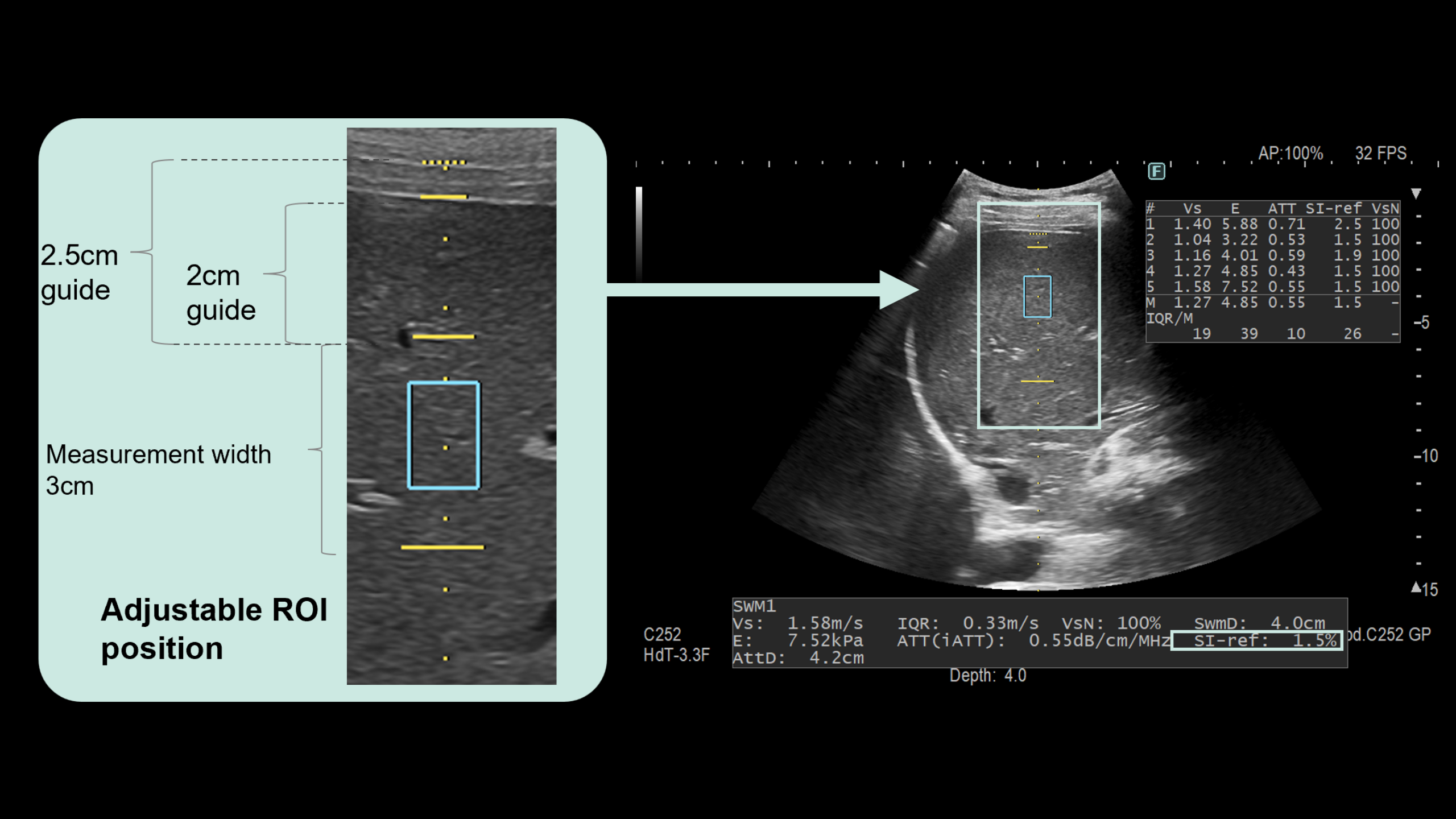
iATT: A cutting-edge tool for precise estimation of hepatic steatosis. By measuring the ATT (Attenuation) measurement, our system offers reliable insights into liver health. Advanced features exclude surface reflections and align ROI depth with WFUMB guidelines. ensuring consistent, accurate, and reproducible results every time.
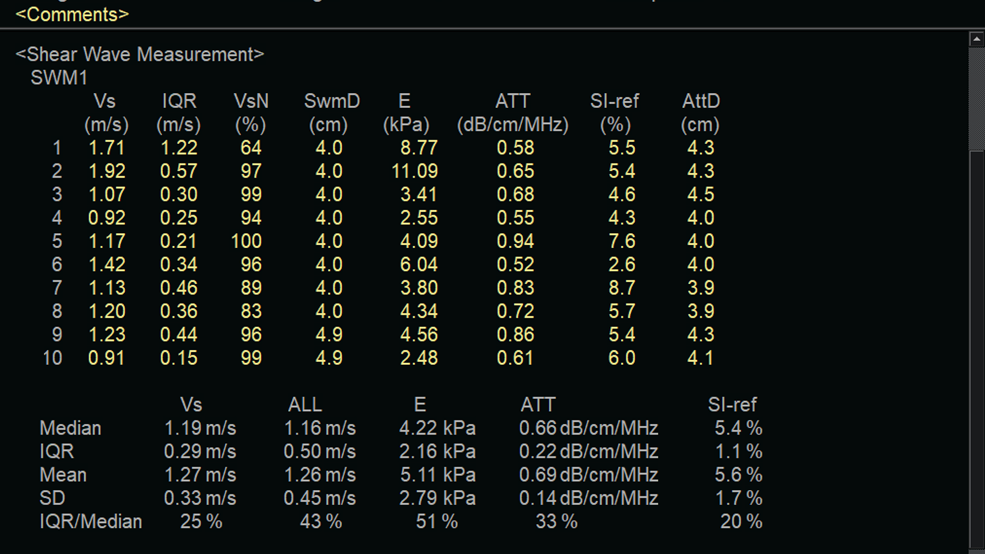
Steatosis Backscatter Index: Introducing a new index for hepatic steatosis. By combining the backscatter coefficient, its variation, and the ATT measurement SBSI delivers more reliable and clinically meaningful assessments. Designed to enhance accuracy and closely align with MRI-PDFF standards, SBSI sets a new benchmark in non-invasive liver health evaluation
Designed to support confident decision-making in routine and advanced liver assessment.
FUJIFILM's Transducer Portfolio
Built For Clinical Demands
DIX is designed to reduce unnecessary interaction—so examinations flow naturally.

Convex/1-6 MHz
C252
Included micro convex shape and features comfortable grips, compact light weight designs and flexible cables.

Micro convex / 1-6 MHz
C23
Micro convex transducer delivering high image quality, great operability and easy application in routine examination, or for difficult access and interventional procedure.

Micro Convex
C421
The unique high frequency micro convex transducer offer reliable performance with an ergonomic easy-to-scan design for inflammatory bowels disease.

Hockey stick - 3-25MHz
L52H
Delivers high image quality in various areas especially for MSK examination.

Linear, 5-13 MHz
L55
Linear transducers with a wide frequency bandwidth provide high-quality images for breast.

Sector / 1-5MHz
S121
The 2D transducers provide precise evaluation of cardiac function.
See more by combining modalities
Fusion imaging and interventional support
See more by combining modalities
DIx enables real-time fusion of ultrasound with CT, MR, and other imaging data—supporting lesion localization, treatment planning, and intervention guidance.
With automated alignment and anatomical recognition, fusion imaging becomes:
- Faster to set up
- Easier to use
- More clinically practical
Real-time fusion
Synchronize ultrasound imaging with CT, MR and other imaging data in real time to strengthen spatial understanding and confidence during complex cases.
Combine modalities
Integrate ultrasound with CT, MR, and other imaging data to bring richer context into the live exam—when visibility and spatial reference matter most.
Lesion localization
Support confident targeting by keeping real-time ultrasound aligned with reference imaging—especially when lesions are subtle or difficult to visualize.
Treatment planning
Planning becomes clearer when Real-time ultrasound is aligned with prior imaging—supporting approach, anatomy, and target location with stronger context.
Intervention guidance
Maintain orientation and consistent visualization throughout the procedure—supporting confident guidance from planning to action.
Empowering knowledge for impactful education
Anatomical information is always valuable for enhancing understanding. Real-time fusion imaging enables simultaneous display of ultrasound alongside other imaging modalities
Important information
Product availability and features may vary by region. Automated measurements require confirmation by a qualified examiner.Specifications subject to change pending regulatory approval.
Built for today—and what comes next
Get in Touch
DeepInsight x is supported by enhanced system performance, faster startup